?️ Proteins (FREE TO VIEW)

The A Level Biologist - Your Hub February 24, 2020
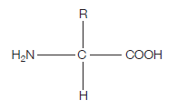
Proteins are at the heart of living organisms. Their functions are very varied, from the hair on your head, to the haemoglobin in your red blood cells (which carries oxygen around the body), to the claws of a lion, to insulin (blood glucose regulation). All these highly varied proteins are made of their building blocks – amino acids. This is what the generalised structure of an amino acid looks like (make sure you can draw this):
If you’re wondering what this actually is, read on. The clues are in the name (as they usually are).
AMINO – the H2N on the left hand side is an amino group
ACID – the COOH on the right hand side is a carboxylic acid group (simply an acid)
The hydrogen (H) on the bottom is there all the time (just like the amino group and the acid group), while the R group is the variable which determines what particular amino acid this will be. For example, if the R group was a hydrogen, the amino acid would be glycine.
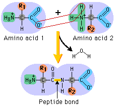
The next diagram shows condensation, and the subsequent formation of a bond between two amino acids (any two). This bond is a peptide bond. The resulting molecule is called a polypeptide.
This video is an excellent tool for understanding the processes by which these amino acids end up in highly structured, complex proteins with varied and important functions within organisms:
The theme of protein structure versus function is really strongly played on in exams, throughout A level biology. The core idea must be learnt, and this is it:
Proteins have a primary, secondary, tertiary and (some only) quaternary structure. The tertiary structure of proteins is their 3D shape which is highly folded and has a unique structure. This structure gives proteins their specific function. For example, if insulin was misfolded, it would cease to function properly. Of course though, the origin of misfolding is likely to be in the primary structure, due to a mutation.
For example, if the gene responsible for coding the amino acid sequence for insulin was mutated, then the insulin’s primary structure (which is the string of amino acids) would be different, leading to a different secondary structure, tertiary structure, and ultimately, a lack of proper function.
NB: The tertiary structure of proteins determines their proper function.
Proteins have many types of bonds in addition to peptide bonds, operating at their different levels of complexity. One of these is ionic bonding which takes place between a positive ion (e.g. NH3+) which donates one or more electrons, and a negative ion (e.g. O-) which accepts them.
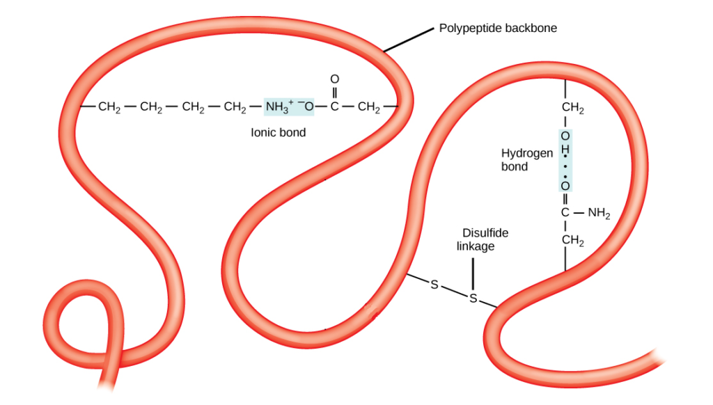
Ionic bonds are weaker than peptide bonds, but stronger than hydrogen bonds. These are momentary bonds between the partial negative charge of an oxygen atom in relation to an available partial positive charge of a hydrogen atom.
Hydrogen bonds are the weakest, while disulfide linkages, bonds or bridges are the strongest. They are covalent bonds between sulphur atoms. Covalent bonds involve a sharing of electrons rather than exchange like ionic bonds.
These bonds are key to the maintenance and formation of a protein’s specific three-dimensional structure. In turn, the structure determines function.
Collagen and Haemoglobin
There are two classes of protein: fibrous and globular based on their structure.
Fibrous proteins don’t usually have a tertiary structure at all, simply forming parallel chains of polypeptides, often cross-linked at intervals to maintain a greater overall arrangement that serves in the structure and support of various tissues including hair, nails and collagen. They are mostly not soluble in water.
Globular proteins on the other hand have a tertiary and sometimes a quaternary structure, are spherical hence the name of globular, normally are water soluble and serve in metabolism such as carrying oxygen in the blood like haemoglobin.
Collagen
Collagen provides support in skin, bones, teeth, tendons and more.
It is very strong and relies on hydrogen bonding to keep its three polypeptide chains together.
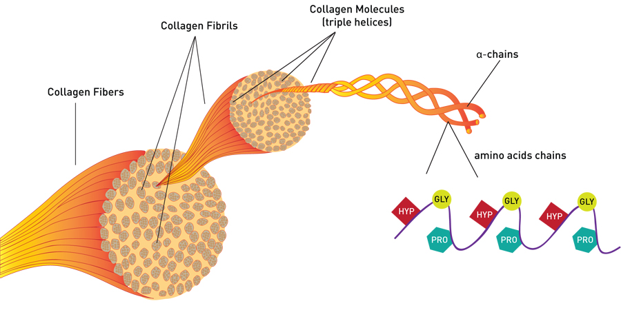
The polypeptide chains are made of hydroxyproline, glycine and proline which are different amino acids.
Haemoglobin
Haemoglobin carries up to 4 oxygen atoms in red blood cells, unloading them in oxygen-poor cells as needed, and replenishing them from the air we breathe, in the lungs.
The extend of its loading with oxygen determines its red colour which otherwise is purple.
The quaternary structure of haemoglobin involves iron ions at its core, 4 of them for each oxygen available for loading. These are called haem groups.
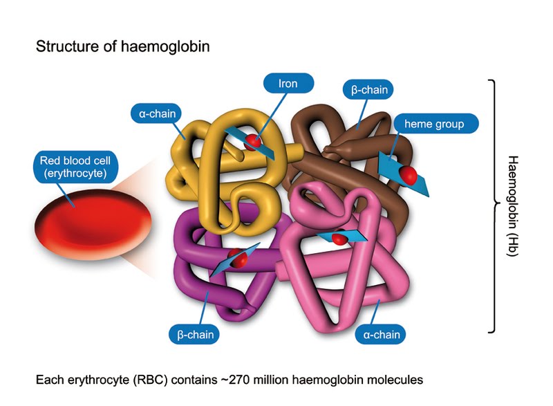
The haem groups are each contained at the centre of 4 polypeptide chains, 2 alpha chains and 2 beta chains.
Ok byeeeeeeeeeeeeee